Multiplexed Fluorescence Detection with the SPECTRA X Light Engine
Lumencor’s SPECTRA X Light Engine has been a leading choice in fluorescence microscopy for over a decade, recognized for its versatility and superior performance (1). It offers researchers exceptional control over excitation spectra, optimizing excitation efficiency while minimizing crosstalk, bleed-through, autofluorescence, and other sources of deleterious background (2). In its new incarnation, the SPECTRA X Light Engine (2023) maintains its user-exchangeable bandpass filters while introducing several significant improvements:
- Expanded Spectral Content: The new model features an increased spectral range with solid-state LED light sources, enhancing compatibility with bandpass filters and fluorophores. New excitation windows at 365 nm and 660 nm are included (Figure 1).
- Greater Output Power: Filtered output power is 100–700 mW for each of the 6 solid-state light sources (previously 50–500 mW).
- Simplified Design: The design now covers the entire spectrum from 365 nm to 750 nm, with teal and near-infrared light sources included as standard (Figure 1).
- Precision Control: Linear output power-to-intensity control setting relationship for all 6 solid-state light sources.
- Improved Safety: Interlocks and design ensure user safety and mitigate the risk of inadvertent light exposure.
The SPECTRA X Light Engine's spectral output and flexibility make it particularly well-suited for fluorescence microscopy and multiplex immunohistochemistry (IHC), a technique used to simultaneously detect multiple markers within a single sample (3, 4). When combined with multiplex IHC, the SPECTRA X enables researchers to gain a more detailed and comprehensive understanding of tissues and cellular environments (5).
The SPECTRA X’s versatile illumination, paired with carefully selected optical filters—such as Kromnigon’s SpectraSplit 7 filter set—and appropriate secondary antibody labeling reagents, ensures a high signal-to-noise ratio with minimal crosstalk and nonspecific labeling. The SpectraSplit 7 filter set allows users to select from a diverse range of fluorochromes, facilitating the customization of a 7-color IHC setup while avoiding spectral overlap (Figure 2). Notably, the often-challenging discrimination of CF594 and Cy5 dyes (Figure 2) is facilitated by the newly extended red (R) wavelength output range of the SPECTRA X (Figure 1). This capacity for optimization is crucial for effective multiplex imaging, making the SPECTRA X a versatile and dependable illumination source for advanced fluorescence microscopy applications.
At Lumencor we embrace continuous product improvement and strive to deliver truly state-of-the-art technical lighting. To that end this next generation SPECTRA X offers enhanced spectral range, increased output power, compact footprint, and precision control in a way no traditional optical bench can. We wonder if your research wouldn’t benefit from this added functionality?
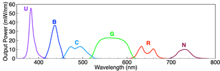
Figure 1. SPECTRA X Light Engine unfiltered spectral output. Letters above the peaks correspond to the six independently controlled light sources. U = UV-Violet, B = Blue, C = Cyan-Teal, G = Green-Yellow, R = Red-Far Red, N = Near-infrared.
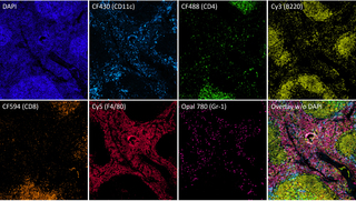
Figure 2. Seven-color imaging of a fixed mouse spleen tissue section at 10X magnification. The tissue was stained with Tyramide Signal Amplification using Kromnigon’s StreptaClick-HRP Multiplex IHC kit and imaged with Kromnigon’s SpectraSplit 7 filter set at 10X magnification. Multiplexed detection of DAPI, CF430, CF488, Cy3, CF594, Cy5 and Opal 780. Biotinylated antibodies used in combination with the fluorochromes include anti-CD11c, anti-CD4, anti-B220, anti-CD8a, anti-F4/80 and anti-Gr-1. Image and supporting technical details provided courtesy of Per Fogelstrand (Kromnigon, Gothenburg, Sweden).
- Aug 20, 2024
- (1) Lumencor’s application bibliography documenting more than 100 published applications of the SPECTRA X Light engine.
- (2) “Bleedthrough or Crosstalk?”
- (3) “The Ever-Expanding Palette of Colors for Multiplexed Fluorescence Detection” – Electro Optics(opens in new window)
- (4) “White Paper: Solid-State Illumination for Multiplexed Fluorescence Detection”
- (5) “Six-Color Imaging with the SPECTRA X Light Engine”