Six-Color Imaging with the SPECTRA X Light Engine
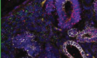
In mammalian tissues, fluorescence excitation in the near infrared (nIR) wavelength region (>650 nm) is useful to avoid the confounding effects of visible-range autofluorescence. The image shown here, generously provided by Dr. Matt Kofron (Cincinnati Children’s Hospital Medical Center), exemplifies this advantage by utilizing excitation light from a Lumencor Light Engine. The specimen is a human kidney section, imaged at 20X magnification. The tissue exhibits high levels of autofluorescence from red blood cells excited at 445 nm, which is pseudocolored yellow in this image. White represents cytokeratin immunodetection using a secondary antibody labeled with the near-infrared fluorophore Alexa Fluor 750. Nucleii stained with DAPI are shown in blue.
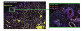
Expanding the spectral range into the near-infrared also increases the number of targets that can be simultaneously detected by multicolor fluorescence imaging. A total of 5 targets plus autofluorescence are present in the image, making use of the full spectra range of the SPECTRA X Light Engine. Three of the targets are mRNA transcripts detected by single-molecule fluorescence in situ hybridization (smFISH) using the fluorophores Opal 520, Opal 570 and Opal 650. These are more clearly evident in an enlargement of the segment marked by the orange rectangle.
TECHNICAL DETAILS
- Specimen: Human kidney section
- Microscope: Nikon Ti2 controlled by NIS Elements software
- Image acquisition: 20X magnification, 5 x 2 tile scan. Pixel size ~ 0.31μm
- Light source: SPECTRA X Light Engine with near-infrared source option
- Andor Zyla 4.2 PLUS camera