Optimizing CELESTA Light Engine Output to Improve MERFISH Workflows
Development of bioanalytical tools to better understand the complexity of tissues has generated a wide array of spatial ‘-omics’ applications. The goal of these techniques is to elucidate how individual cells function within the spatial context of a complex tissue environment. These techniques have been used to build defined tissue atlases to better understand disease states and development (1). Multiplexed error-robust fluorescence in situ hybridization (MERFISH) is a spatial technique that has been developed to capture transcriptomic information at the single-cell level. MERFISH utilizes multiple rounds of fluorescent RNA probe hybridization to barcode-specific RNA species. This allows for the identification of gene expression patterns and cell-type identification within tissues. MERFISH and other spatial ‘-omics’ techniques require specialized imaging workflows, and face limitations to imaging speed and data quality.
Dr. Meng Zhang, Assistant Professor Molecular & Cellular Biology at Scripps Research Institute, is developing improved MERFISH workflows. Dr. Zhang aims to better understand the fundamental mechanisms of tissue cohesion and function (2). The primary limitations to current MERFISH workflows have been light output power and output stability. These limitations affect researchers’ ability to detect low abundance transcripts and the rate of photobleaching of fluorescent probes. Since replicating MERFISH experiments are costly and time consuming, optimizing light output conditions is a key part of advancing MERFISH workflows. Having worked with a CELESTA Light Engine for MERFISH under Professor Xiaowei Zhuang at Harvard University, Dr. Zhang approached Lumencor Inc. to further enhance her imaging system and improve her MERFISH workflows.
Working with the team at Lumencor Inc., adjustments in optical output power tailored for individual light sources made it possible to meet their application needs. Dr. Zhang and her team saw significant improvements to their MERFISH data, including reduced background and improved sensitivity. These improvements led to more accurate gene profiling and better cell-type identification. As a result of customizing a CELESTA Light Engine for MERFISH, Dr. Zhang and her team are advancing our knowledge of intracellular communication and tissue organization.
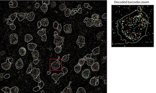
![Figure 1: Decoded MERFISH image in a single field of view taken for a mouse brain slice. Image is shown as a maximum intensity projection. Pixels are color-coded to specific MERFISH barcodes. White boundaries indicate single cells segmented by DAPI for analysis. Scale bars, 20m (left) and 5m (right). Adapted from [2] under CC BY 4.0.](https://cms.lumencor.com/system/uploads/fae/image/asset/1253/xs_2.jpg)
Figure 1: Decoded MERFISH image in a single field of view taken for a mouse brain slice. Image is shown as a maximum intensity projection. Pixels are color-coded to specific MERFISH barcodes. White boundaries indicate single cells segmented by DAPI for analysis. Scale bars, 20μm (left) and 5μm (right). Adapted from (2) under CC BY 4.0.
![Figure 2: Integrated MERFISH and single-cell RNA sequencing data used to create a mouse whole-brain cell atlas. (Left) Uniform manifold approximation and projection (UMAP) of integrated MERFISH and scRNA-seq data of cell subclass identification based on gene expression. (Right) Spatial maps of cell subclasses in representative mouse brain slices. Scale bar=1mm. Adapted from [3] under CC BY 4.0.](https://cms.lumencor.com/system/uploads/fae/image/asset/1254/xs_3.jpg)
Figure 2: Integrated MERFISH and single-cell RNA sequencing data used to create a mouse whole-brain cell atlas. (Left) Uniform manifold approximation and projection (UMAP) of integrated MERFISH and scRNA-seq data of cell subclass identification based on gene expression. (Right) Spatial maps of cell subclasses in representative mouse brain slices. Scale bar=1mm. Adapted from (3) under CC BY 4.0.

"The CELESTA Light engine is an excellent illumination source for MERFISH imaging with high and stable output power. With its compact and fully integrated design, it's very easy to incorporate into our existing widefield or confocal MERFISH imaging platforms. I appreciate its robustness and long-term stability."
-Dr. Meng Zhang

- Apr 22, 2025
- (1) Z Yao, CTJ van Velthoven, M Kunst, et al. A high-resolution transcriptomic and spatial atlas of cell types in the whole mouse brain. Nature (2023) 624(7991):317-332.(opens in new window)
- (2) M Zhang, SW Eichhorn, B Zingg, et al. Spatially resolved cell atlas of the mouse primary motor cortex by MERFISH. Nature (2021) 598:137-143.(opens in new window)
- (3) M Zhang, X Pan, W Jung et al. Molecularly defined and spatially resolved cell atlas of the whole mouse brain. Nature (2023) 624(7991):343-354.(opens in new window)
- (4) Zhang, M. et al. Molecularly defined and spatially resolved cell atlas of the whole mouse brain. Nature 624, 343-354 (2023)(opens in new window)