UTHealth Confocal Imaging Workshop
The Center for Advanced Microscopy at UTHealth at Houston, a Nikon Center of Excellence, is a state-of-the-art microscopy facility serving the Texas Medical Center. We jointly hosted a full house at the recent confocal imaging workshop.
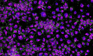
Attendees interrogated their own samples utilizing Lumencor’s ZIVA Light Engine a Yokogawa CSU, and a Nikon Ti2 microscope. Judge the image quality, success of the hardware integration and user experience for yourself.
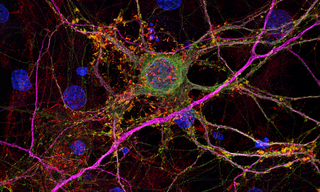
Figure 1. Rodent Neuron Cells

"What took me 2 hours to image on my Zeiss laser scanning confocal microscope only took 2 minutes today!?"
—Amaya Craft
Student in Genetics and Epigenetics, UTHealth

"Wow, this system was so much faster and easier to use compared to our current setup. We loved the beautiful images that we took on this system. The 3D rendering made possible via this scope allowed us to view protein localization in a much more accurate and precise manner, contributing strongly to our research."
—Michael Claxton, Ph.D.
Postdoctoral Fellow, Houston Methodist Hospital

"The high spatial and temporal resolution of real-time videos, acquired using the “ZIVA x Yokogawa CSU-W1 equipped with SoRA”, is sufficient to resolve the dynamic membrane interactions between T-cells and cancer cells and to visualize subsequent T-cell-induced cancer cell death.”
—Yunfei Wen, Ph.D.
Associate Professor, University of Texas MD Anderson Cancer Center

- Jul 31, 2025