Highlighting smFISH with SPECTRA | Lumencor FISH Light Sources
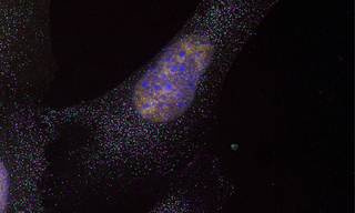
1st Place - George McNamara and Lauren Blake
5plex mouse embryonic fibroblasts expressing MS2-tagged beta-actin mRNA. Red Halo-JF549-NLS-MCP, Green Atto594 POLR2A mRNA FISH, Blue DAPI, Cyan Cy5 beta-actin-MS2 mRNA FISH, Magenta Alexa Fluor 488-anti-DDX6 immunofluorescence. Microscope details at http://confocal.jhu.edu/current-equipment/fishscope. Specimen preparation by Lauren Blake, Prof. Bin Wu’s lab (JHU Biophysics). Imaging by George McNamara, Ross Fluorescence Imaging Center.
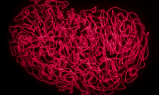
2nd Place - Ariel Waldman
A nostoc discovered in Antarctica autofluorescing under TRITC excitation. This nostoc was found inside a microbial mat in the Dry Valleys of Antarctica. A nostoc is a genus of cyanobacteria with beaded filaments intricately woven inside a gelatinous pouch. This image highlights the structure of the beaded filaments and their scaffolding within the gelatinous pouch.
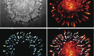
3rd Place - Tejeshwar Rao
Cos-7 cells spread on a tension gauge tether (TGT) surface and imaged on a Nikon Ti2 eclipse microscope using a Lumencor light engine and a turret wheel with different excitation and emission filter cubes - The RICM image (panel 1) was taken by removing the emission filter from the path. Cell tension indicated by TGT probe opening (panel 2) and paxillin staining (panel 3).
- Sep 21, 2020