Does your High-Throughput Screening workflow demand 2D and 3D Cell Models? Advance your Cardiac Research with the VOLTA Scanner
Human induced pluripotent stem cell-derived cardiomyocytes (hIPSC-CMs) are transforming the preclinical assessment of cardiac safety. Whether in 2D monolayers or 3D cardioids with supporting cardiac fibroblasts and endothelial cells, hIPSC-CMs are powerful tools for evaluating drug-induced cardiotoxicity and electrophysiological effects (Figure 1). While 2D models offer relative simplicity and ease-of-use, 3D cardioids more closely model human cardiac physiology [1, 2]. Together, these models provide complementary insights, enhancing the depth and translational relevance of cardiac studies. Now, both 2D and 3D models can be studied on a single, highly-integrated, turnkey platform.
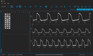
Lumencor’s VOLTA Scanner is a kinetic optical plate reader, designed to support high-throughput screening of both 2D and 3D hIPSC-CMs (Figure 2) [3]. It generates traditional electrophysiological data that is associated with traditional patchclamp studies, however VOLTA uses optical transduction for signal generation. Whether assessing cellular functional responses to test compounds or comparing CM performance, VOLTA enables consistent, reliable, scalable data acquisition—all on the same platform. VOLTA supports:
- Live-Cell Optophysiology Studies: Optically reports on cellular physiology using fluorescence labels and living cells, preserving cellular function throughout cell lifetimes.
- Dual-Model Compatibility: Probes both 2D and 3D cell models in drug screens and toxicity studies for more comprehensive functional insights than a single model can offer.
- Fast, Non-Disruptive Scanning: Captures data from 96- and 384-well plates within two minutes while preserving sample health and data quality.
- Robust Workflow: Streamlined hardware and software enable automated, high-throughput analyses with well-behaved, reproducible results and push button operation.
- Exceptional Temporal Resolution: A 10 kHz sampling rate detects subtle, time-dependent changes in transmembrane action potential [4], even in complex 3D structures.
Whether you're investigating cardiac toxicity, evaluating compound efficacy, or validating novel disease models, the VOLTA Scanner provides the flexibility and precision needed for modern cardiac research.
VOLTA Scanner can illuminate your understanding of 2D and 3D cellular models in one powerful, optophysiology platform. Could your screens benefit from a turnkey, optical plate reader with exceptional reproducibility, speed and precision?
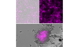
Figure 1. 2D (top) and 3D (bottom) hIPSC-cardiomyocytes labeled with BeRST voltage sensitive dye. CMs imaged at 20x or 10x magnification, respectively. Scale bar applies to bottom image.

Figure 2. VOLTA software displays fluorescence waveforms of 2D and 3D hIPSC-CMs collected on the VOLTA Scanner at 10 kHz sampling rate over time. Fluorescence of BeRST voltage sensitive dye excited by 660 nm light reporting changes in transmembrane action potential for 2D (well B02) and 3D (wells C02 and C03) hIPSC-CMs at two cell densities (10,000 cells / C02; 20,000 cells / C03).
- Jun 05, 2025
- KD Shead, E Huethorst, GL Smith et al. (2024) JACC. CardioOnco, 6(5), 678-683.(opens in new window)
- S Stoter, A Maria et al. (2020) Clinical Therapeutics 42(10), 1892-1910(opens in new window)
- VOLTA Scanner: A Turnkey, Optical Kinetic Plate Reader for Cardiac Safety Testing
- VOLTA Scanner: High-Throughput Cardiomyocyte Electrophysiology with an Optical Pacemaker