MIRA Light Engine for Microscopy at VT's Neuroscience School
MIRA on the Brain
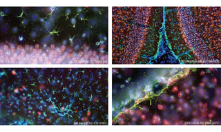
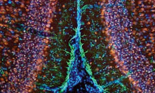
At Virginia Tech’s School of Neuroscience, Professor Ian Kimbrough provides students with hands-on experience of real-world neuroscience research techniques including immunohistochemistry, neuroanatomy, and microsurgery, just to name a few. Fluorescence microscopy is the fundamental and essential tool underpinning these techniques. Professor Kimbrough’s teaching laboratory has a fleet of ~20 Nikon Eclipse E200 fluorescence microscopes equipped with Lumencor MIRA Light Engines. Solid-state illumination with the MIRA Light Engine provides powerful, stable, and responsive fluorescence excitation for visualization of coronal mouse slices to elucidate cellular definition of mouse brain anatomy. The images below are representative of many images obtained by the 2018 cohort of Professor Kimbrough’s neuroscience laboratory class using the MIRA Light Engine. A video presentation including more images can be viewed here.. The coronal mouse brain sections were stained using immunohistochemical techniques to fluorescently label various brain cell types and structures: neuronal nuclei (blue), neurons (red), astrocytes (green), and NISSL bodies (yellow).