Validation of mammalian cell surface display of SARS-CoV-2 spike protein using the SOLA Light Engine
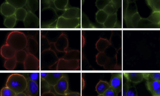
The SARS-CoV-2 spike glycoprotein is the immunogen encoded by mRNA vaccines against COVID-19, and a target for neutralizing monoclonal antibody therapeutics. Recognizing the current need for high-throughput determination of expression levels, receptor binding, and antibody escape across hundreds of spike glycoprotein variants, a team of researchers from the University of Texas recently reported the development of Spike Display technology (1). Spike Display enables transgenic expression of spike glycoproteins on the surface of mammalian cells. Spikes are expressed with appended 3X FLAG tags and protease cleavage and affinity purification sites. As part of the platform validation, two-color immunofluorescence microscopy using a SOLA Light Engine to confirm cell membrane localization of SARS-CoV-2 spikes and binding of proteins ACE2, REGN10933 and 4A8 (Figure 1). ACE2 (angiotensin-converting enzyme 2) is a key intermediary in the viral infection process. REGN10933 (casirivimab) is one of the two components of Regeneron’s REGEN-COV neutralizing antibody cocktail. 4A8 is a neutralizing antibody (nAb) targeting the NTD (N-terminal domain) of the spike protein, as opposed to the receptor binding domain targeted by ACE2 and REGN10933. Using Spike Display, the team assayed ~200 SARS-CoV-2 spike variants for expression level, ACE2 binding, and recognition by neutralizing antibodies, demonstrating its potential for accelerating vaccine design and rapidly assessing the effects of mutations in emerging viral variants.
![Figure 1. Immunofluorescence microscopy validation of SARS-CoV-2 spike glycoprotein localization on human embryonic kidney (HEK293T) cell surfaces. Images acquired using 60X magnification on a NikonTs2R-FL microscope with illumination from a SOLA Light Engine. Green fluorescence indicates expression level. Red fluorescence indicates binding of ACE2 and neutralizing antibodies REGN10933 and 4A8. Blue fluorescence indicates nuclear staining by Hoechst 33342. Reproduced from [1] under CC BY 4.0.](https://cms.lumencor.com/system/uploads/fae/image/asset/797/xs_WebsiteImage.jpg)
Figure 1. Immunofluorescence microscopy validation of SARS-CoV-2 spike glycoprotein localization on human embryonic kidney (HEK293T) cell surfaces. Images acquired using 60X magnification on a NikonTs2R-FL microscope with illumination from a SOLA Light Engine. Green fluorescence indicates expression level. Red fluorescence indicates binding of ACE2 and neutralizing antibodies REGN10933 and 4A8. Blue fluorescence indicates nuclear staining by Hoechst 33342. Reproduced from [1] under CC BY 4.0.
- Jan 10, 2022