Cell Painting is a multiplexed imaging assay introduced by Gustafsdottir and colleagues in 2013. As originally envisioned, six chemical stains are used to fluorescently label and image eight distinct cellular constituents: DNA, cytoplasmic RNA, nucleoli, actin, Golgi apparatus, plasma membrane, endoplasmic reticulum, and mitochondria. The assay provides insights into the mechanisms of action underlying phenotypic changes and has been widely adopted in drug discovery and preclinical safety pharmacology (Gustafsdottir et al., 2013. PLoS ONE, 8:e8099).
As the drug discovery community has demanded improvements to throughput and fast acquisition of time-course data from single microwell sample plates, the technique continues to evolve and mature. Increasingly high-resolution Z-stacked brightfield imaging and in silico processing as a complement to traditional, fluorescence multiplexed assays for Cell Painting is being developed and adopted. For example, researchers at the University of Cambridge and AstraZeneca (Cambridge, UK) demonstrated that machine learning algorithms can generate Cell Painting in the form of level insights from merely three optical slices in a Z-stack combined with label-free brightfield imaging (Cross-Zamirski et al., 2022. Sci Rep, 12:10001). More recently, the research team at Recursion Pharmaceuticals, Inc.'s (Salt Lake City, UT) demonstrated improvements to machine learning algorithms produce image analysis quality that is up to 95% equivalent to fluorescence Cell Painting results (Baker et al. 2024). The costs associated with fluorescence Cell Painting are known and high; including but not limited to expensive fluorescent reagents, extensive staining time (often three hours or more), the destructive nature of repeated staining and stripping steps in more highly ordered multiplexed fluorescence protocols, large file sizes from five-plex imaging and higher, and the single-use nature of fixed and permeabilized samples. Brightfield imaging presents a more efficient, scalable, and cost-effective alternative.
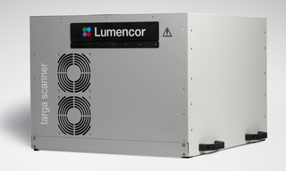
To address the hardware imaging needs for brightfield-based Cell Paining Lumencor has developed the TARGA Imager for high-throughput screening of multi-well plates. TARGA’s workflow includes laser-based autofocus on every well, acquisition of a three-slice Z-stack centered on the autofocus plane, and high-resolution brightfield imaging. The resulting 16-bit TIFF image stacks can be analyzed with machine learning algorithms to achieve mechanistic profiling performance comparable to Cell Painting. TARGA’s data competes with more complex, destructive, costly fluorescence Cell Paining while eliminating the need for complex fluorescent staining and extended sample preparation and destruction. Moreover, TARGA is fast: TARGA can image a 1536-well plate in under two minutes. This represents a 120-fold improvement in time-to-data compared to conventional five-color Cell Painting workflows (2 minutes using TARGA vs. typical 240 minutes for high-content Cell Painting; Cimini et al., 2023. Nat Protoc, 18(7):1981–2013). Additionally, because fewer images are required compared to fluorescence Cell Painting, the downstream data storage requirements are significantly reduced 16-fold (11.52 GB vs 192 GB).

- Jul 15, 2025

- B. Cimini and colleagues. Nat Protoc. 2023. 18(7): 1981-2013. “Optimizing the Cell Painting assay for image-based profiling.”(opens in new window)
- C. Baker and colleagues. “How Phenomic foundation models are empowering the brightfield comeback.”(opens in new window)
- J. Cross-Zamirski and colleagues. Scientific Reports. 2022. 12: 10001. “Label-free prediction of cell painting from brightfield images.”(opens in new window)
- M. Bray and colleagues. Nat Protoc. 2016. 11: 1757-1774. “Cell Painting, a high-content image-based assay for morphological profiling using multiplexed fluorescent dyes.”(opens in new window)
- S. Gustafsdottir and colleagues. PLoS ONE. 2013. 8:e80999. “Multiplex cytological profiling assay to measure diverse cellular states.”(opens in new window)


