MERFISH Imaging of Gene Expression with CELESTA Light Engine
MERFISH (multiplex error robust fluorescence in situ hybridization) is an imaging technique that profiles cell populations based on the identification of thousands of RNA transcripts per cell. The CELESTA Light Engine is an ideal and widely-adopted solid-state illumination source for this application. In a recent paper published in Nature, Wheeler and co-workers used MERFISH imaging with a CELESTA Light Engine to quantify the expression of nine specific astrocyte and T-cell markers[1]. Five of the CELESTA Light Engine’s seven laser lines were used in the highly multiplexed MERFISH imaging protocol. The overall objective of the research described in the paper was to characterize astrocyte populations that contribute to pathogenesis in a preclinical model of multiple sclerosis.
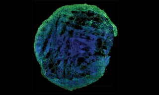
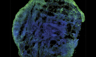
Brain Organoid
50 micro thick slice cut from a brain organoid structure derived from induced pluripotent stem cells
Microscope: Nikon Ti2
Objective: Nikon 60X oil Plan Apo Lambda, NA 1.4
SD confocal module: Crest X-Light V3
Confocal Laser bank: Lumencor CELESTA Light Engine
Staining:
- Magenta: TBR1 stained with Alexa 750. TBR1 is a transcription factor important for the differentiation of the nuclear glutamatergic neurons during cortical development. Staining specific for layer VI neurons.
- Green: MAP2 stained with Alexa 488. MAP2 is a protein associated with the microtubules present in the cytoskeleton of the neuronal dendrites.
- Blue: DAPI
- Sep 30, 2021