Lumencor Light Engines for Microscopy at VT’s Neuroscience Centers
At Virginia Tech’s School of Neuroscience, Professor Ian Kimbrough provides students with hands-on experience of real-world neuroscience research techniques including immunohistochemistry, neuroanatomy, and microsurgery, just to name a few. Fluorescence microscopy is the fundamental and essential tool underpinning these techniques.
Ian Kimbrough, an assistant professor in the School of Neuroscience, a part of the College of Science, came to Virginia Tech in 2016. He specializes in research using live in vivo imaging to model neurological diseases, such as brain tumors and Alzheimer’s. Before becoming a neuroscientist, Kimbrough specialized in digital media design. He has now combined the two specialties. He uses laser scanning microscopes and has developed novel imaging techniques to study brain diseases. Moreover his students benefit from the wealth of knowledge and experience he brings to the classroom.
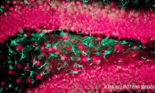
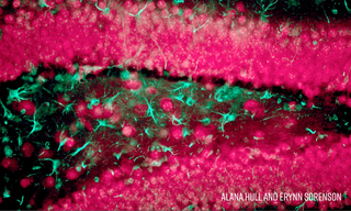
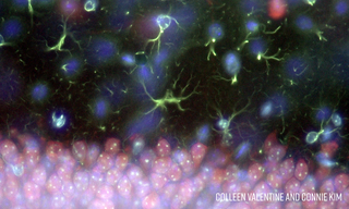
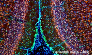
Professor Kimbrough’s teaching laboratory has a fleet of ~20 Nikon Eclipse E200 fluorescence microscopes equipped with Lumencor MIRA Light Engines. Solid-state illumination with the MIRA Light Engine provides powerful, stable, and responsive fluorescence excitation for visualization of coronal mouse slices to elucidate cellular definition of mouse brain anatomy. The images below are representative of many images obtained by the 2018 cohort of Professor Kimbrough’s neuroscience laboratory class using the MIRA Light Engine. A video presentation including more images can be viewed here. The coronal mouse brain sections were stained using immunohistochemical techniques to fluorescently label various brain cell types and structures: neuronal nuclei (blue), neurons (red), astrocytes (green), and NISSL bodies (yellow).

- Aug 15, 2023