Imaging Algae for Water Quality with SOLA Light Engine
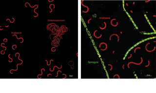
Cyanobacteria, also referred to as blue-green algae, form dense and sometimes toxic blooms in freshwater and marine environments. Such blue-green algae threaten ecosystem function and degrade water quality for recreation, drinking water, fisheries, and human health (1). When characterizing the composition of algal blooms, fluorescence microscopy is superior to DIC phase microscopy (2). A rapid one minute fluorescent technique can be used to generate a composite image of freshwater cyanobacteria and green algae, based on the absorption characteristics of their unique pigments. Green algae primarily contain chlorophyll and are excited efficiently with blue light. Cyanobacteria contain phycobilisomes, which are best excited with green light. Pseudo-colored images derived from each of the wavelength excitations can be generated to distinguish algae from cyanobacteria. The pseudo-colored images below are of water samples drawn in December 2021 from Lake Betz, North Carolina. They were acquired using a SOLA Light Engine by Dr. Robert Zucker and Emma Brentjens at the Center for Public Health and Environmental Assessment at the U.S. Environmental Protection Agency, Research Triangle Park, North Carolina.
- May 10, 2022
- (1) J Huisman, GA Codd, PM Visser er al. (2018) Cyanobacterial blooms. Nat Rev Microbiol 16:471-483(opens in new window)
- (2) Schulze K, López DA, Tillich UM, Frohme M. (2011) A simple viability analysis for unicellular cyanobacteria using a new autofluorescence assay, automated microscopy, and ImageJ. BMC Biotechnol. 11:118.(opens in new window)