Many life science applications benefit from the brilliance, spectral content and temporal control of light output provided by Lumencor Light Engines. A glance at some recent research publications gives a perspective of the diversity of these applications.
Live Cell Imaging
Direct observation of protein complexes and membranes in living cells provides information on how they interact and when and where they are involved in the process of autophagy. Researchers from the Babraham Institute (Cambridge, UK) provide detailed protocols for live cell time-lapse imaging of three protein participants in the autophagy pathway, DFCP1, ATG13, and LC3, tagged with GFP, mCherry, and CFP, respectively. Excitation for CFP (434/17 nm), GFP (488/10 nm) and mCherry (560/25 nm) is supplied by a SPECTRA X Light Engine. Correlative super resolution images from cells that have previously been imaged live reveal proteins or membrane assemblies that make a dynamic contribution to autophagosome formation but fall below the resolution limit of wide-field light microscopy.
Genomic Analysis
Rapid and accurate profiling of pathogen genotypes in complex samples remains a challenge for existing molecular detection technologies. A team from the University of California San Diego recently described a digital high resolution melting (dHRM) analyzer for identification of bacterial pathogen DNA sequences. The analyzer performs 20,000 simultaneous reactions comprising digital PCR followed by HRM sequence fingerprinting on a microfluidic chip.On-chip thermal control and fluorescence detection using a SPECTRA X Light Engine produce temperature-registered two-color fluorescence images of the chip that are transformed into 20,000 DNA melt curves by an automated image analysis algorithm.
Neuroscience
Signaling in neuronal axons and dendrites necessitates the maintenance and modification of their local proteomes. Local translation of mRNAs into protein is one solution that neurons use to meet synaptic demand and activity. Imaging mRNA in living cells using the MS2-GFP labeling system provides dynamic information to augment static snapshots obtained using fluorescence in situ hybridization. Moon and Park from Seoul National University describe protocols for imaging GFP-labeled endogenous β-actin mRNA in cultured neurons using a SOLA SE Light Engine controlled by Micro-Manager image acquisition software. They note the importance of using low power, pulsed illumination and high EMCCD camera gain to minimize photobleaching and phototoxicity during image acquisition.
Intravital Imaging
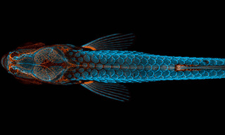
How does hydrogen peroxide (H2O2) act to recruit subcutaneous antimicrobial leukocytes to a superficial injury site? This question has been addressed by imaging the penetration of exogenous H2O2 in wounded zebrafish larvae expressing the fluorescent H2O2 sensor HyPer. Time-lapse fluorescence ratio imaging of HyPer-transgenic zebrafish using the blue (438/57 nm) and cyan (475/28 nm) outputs from a SPECTRA X Light Engine was performed at 1 image pair per minute for 60 minutes. The data indicated that direct redox signaling by wound-derived H2O2 is largely confined to the wound margin.
- Aug 14, 2017

- Biophys J . 2017 May 9;112(9):2011-2018. doi: 10.1016/j.bpj.2017.03.021.(opens in new window)
- Methods Enzymol . 2016;572:51-64. doi: 10.1016/bs.mie.2016.02.015. Epub 2016 Mar 22.(opens in new window)
- Methods Enzymol . 2017;587:1-20. doi: 10.1016/bs.mie.2016.09.050. Epub 2016 Nov 17.(opens in new window)
- Sci Rep . 2017 Feb 8;7:42326. doi: 10.1038/srep42326.(opens in new window)


