Skip Navigation

Microscopy

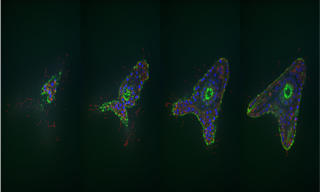
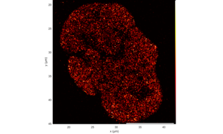
Light microscopy is a core research technique in cell biology. Yet its utility extends far beyond, across all fields of research, manufacturing and testing where structural information on micron-scale features is required. Light microscopy encompasses a multitude of specific techniques, some of which are listed below. Lumencor’s solid-state Light Engines excel in all of them.
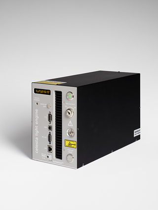
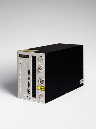
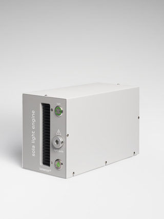
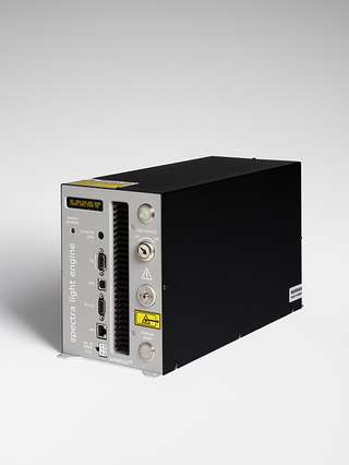
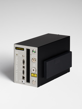
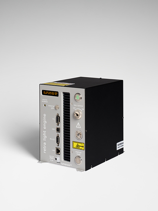
Widefield Fluorescence Microscopy is the least specialized and most common type of fluorescence microscopy. Microscope mercury arc and metal halide light sources, ubiquitous for many years but encumbered by erratic performance, have been largely replaced by high-performance, solid-state Light Engines that are mercury-free, clean, and green. Solid-state light sources are subdivided into white light and color selective output types. White-light sources are direct replacements for mercury arc and metal halide lamps with superior stability, longer operating lifetimes, more responsive control characteristics and lower operating costs. Color-selective Light Engines eliminate the need for mechanical filter switching in multicolor imaging protocols, enabling faster data acquisition.

Confocal Microscopy provides 3-dimensional spatial information by spatially constraining the excitation light. This translates into a requirement for higher initial light intensities compared to widefield microscopy. Consequently, laser light sources are usually preferred over LEDs for confocal microscopy applications.

Super-Resolution Microscopy provides spatial resolution in the 20–200 nm range, beyond the limit of widefield fluorescence microscopy (~200 nm). As with confocal microscopy, spatially constrained excitation light is required and laser light sources are usually preferred.

Transmitted Light Microscopy generally requires lower light intensities than fluorescence microscopy, allowing smaller, passively cooled light sources to be used. Tungsten-halogen lamps, predominant for many years, have been replaced by solid-state microscope light sources, for largely the same reasons that they have supplanted mercury arc lamps in widefield fluorescence microscopy. In particular, the spectral distribution (color temperature) of solid state light sources does not vary with output intensity, an important advantage in terms of consistent color rendition.

Darkfield Microscopy provides an image of the light scattered by the specimen, using spatial filtering to exclude unscattered light. Under darkfield (DF) illumination, flat surfaces appear dark, and features such as cracks, pores and etched boundaries are enhanced. Darkfield epillumination is therefore useful for detection of defects in opaque, unstained materials such as semiconductor wafers. Because the illumination must be spatially filtered, light sources with higher output intensity than those used for transmitted light microscopy are required.